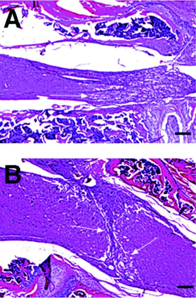
Figure 4.

Representative micrographs illustrating histopathological staining with hematoxylin and eosin. Sagittal section of the spinal cord from a paraplegic control (A) and a representative CM101-treated (B) mouse sacrificed on day 6 after injury after four treatments with 30 μg/kg CM101. Amount of hemorrhage, fibrosis, and gliosis formed in the spinal cord (area between arrows) in the CM101-treated mice is reduced compared with control. (Bars = 100 μm.)
