
Figure 3.
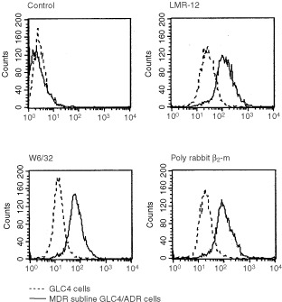
FACS analysis of GLC4 and MDR subline GLC4/ADR cells with control Mab, LMR-12, W6/32 and rabbit polyclonal anti β2-m. The different anti-MHC class I anti sera show similarly positive signals on the GLC4 cells, which are equally increased on the MDR subline GLC4/ADR. Antibody binding was detected using FITC-labelled rabbit-anti-rat serum, -anti-mouse serum or swine-anti-rabbit serum. The samples were analysed on a FACS-Star flow cytometer.