
Figure 4.
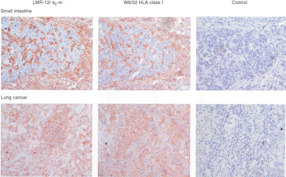
Staining of frozen sections of tumour samples with LMR-12, W6/32 and control Mab. Upper panel: small intestinal cancer samples (showing only staining in the surrounding tissue). Lower panel: lung cancer samples (showing staining in the tumour cells and in the surrounding tissue). In the two panels a similar staining pattern is observed for both LMR-12/ β2-m and W6/32 HLA class I molecules. Colour development was with 0.02% (w v−1) amino-ethyl-carbazole and 0.02% (v v−1) H2O2.