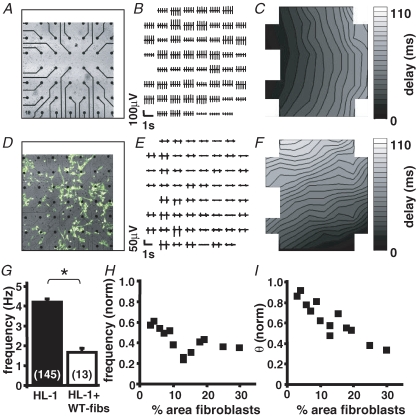
Figure 1. Spontaneous activity and excitation spread in homo- and heterocellular cardiomyocyte preparations:
Representative recordings of a homocellular HL-1 (A–C) and heterocellular (D–F) HL-1 + WT-fibs monolayer Transmission pictures (A and D) display a section of the electrode field and the location of the fibroblasts within the monolayer (white; areas in D). In homo- and heterocellular cultures only preparations were used where all 60 electrodes of the array exhibited synchronized activity (B and E). In both preparations the contour plots (C and F) exhibit continuous excitation spread; however, with a decreased velocity in the heterocellular preparation (homocellular: 2.0 cm s−1; heterocellular: 1.1 cm s−1). G, comparison of the beating frequency recorded in homo- (n = 145) and heterocellular (n = 13) monolayers of HL-1 cells and WT-fibs (*P < 0.05). The beating frequency (H) and conduction velocity (θ) (I) of heterocellular cultures were normalized to those of HL-1 monolayers; both parameter correlate negatively with the percentage area of fibroblasts on the MEA.

