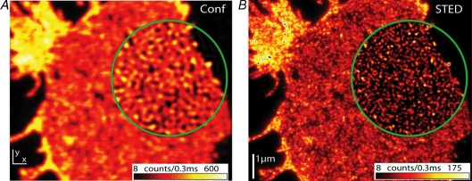
Figure 2. Nanoscale resolution STED microscopic analysis of SNAP-25 organization.
Confocal and STED micrograph of a plasma membrane sheet immunostained for SNAP-25. Encircled regions show linearly deconvolved data. Conventional resolution provided by confocal microscopy (A) is not sufficient to resolve individual SNAP-25 clusters present at high density. STED microscopic resolution (B) reveals that individual clusters are smaller than 60 nm average size (published with permission from Willig et al. 2006; Copyright Institute of Physics Publishing).

