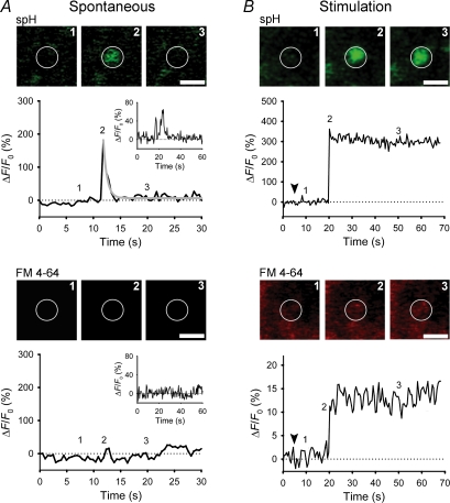
Figure 2. Subnanometer fusion pores in spontaneous peptidergic vesicles.
A and B, sequential images of a single vesicle and changes in spH and FM 4-64 fluorescence at the vesicle site at rest (A) and after stimulation (B). Exocytosis resulted in a rapid increase in spH fluorescence, followed by a rapid exponential decline (grey line; spontaneous) or a persistent elevation (after stimulation). Insets in A show a repetitive spontaneous exocytotic event. Note that stimulation triggered loading of FM 4-64 dye into vesicles probably due to larger pore diameter. Numbers on plots correspond to times when images were recorded. White circles indicate sites of exocytic events. Scale bar represents 1 μm. Arrowheads indicate the onset of stimulation. Modified from Vardjan et al. (2007) with the permission of The Society for Neuroscience.

