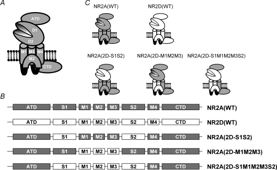
Figure 1. Cartoon illustration of an NMDAR subunit and the various chimeric constructs examined.
A, cartoon sketch of an ionotropic glutamate receptor subunit showing the proposed membrane topology of three membrane spanning domains (M1, M3 and M4) and a re-entrant loop (M2), and the location of the amino terminal domain (ATD) and carboxy terminal domain (CTD). The ligand binding domains (denoted D1 and D2) are formed by the S1 and S2 regions of the protein, which come together to form a hinged clamshell-like structure. B, linear representation of the various NMDAR constructs investigated and the nomenclature used in this study. Regions originating from the NR2A subunit are shown in grey, while those originating from the NR2D subunit are shown in white. C, cartoon representation of these constructs showing how the various functional domains from the NR2D subunit are incorporated into the three chimeric subunits.

