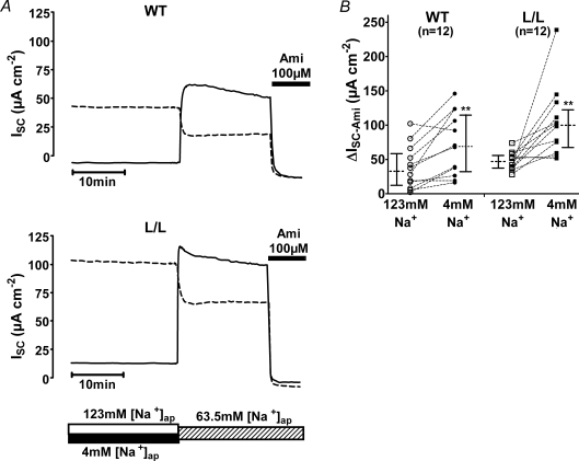
Figure 6. Prolonged exposure of colon tissue to low apical Na+ results in an up-regulation of ENaC which is preserved in Liddle mice.
Representative traces are shown in A for one wild-type animal (WT; top traces) and one L/L animal (bottom traces). Paired colon tissue preparations obtained from the same animal were mounted into Ussing chambers and were exposed either to an apical solution containing 4 mm Na+ (low sodium) or to an apical solution containing 123 mm Na+ (high sodium). The basolateral compartment contained standard mouse Ringer solution. Tissue preparations were maintained under these conditions for 8 h. At the time indicated by the bar below the traces, the apical bath solution was switched to a bath solution containing 63.5 mm NaCl. This was achieved either by replacing 50% of the low sodium bath solution by high sodium bath solution (ISC traces with continuous lines) or by replacing 50% of the high sodium bath solution by low sodium bath solution (ISC traces with dashed lines). Application of 10−4m amiloride (Ami), as indicated by the black bar, revealed the ENaC-mediated ISC component (ΔISC-Ami). B, summary of ΔISC-Ami values obtained in similar experiments as shown in A. Each circle or square represents an individual ΔISC-Ami value measured either in colon tissue pre-incubated in high sodium (123 mm; open symbols) or in low sodium (4 mm; filled symbols), using colon tissue from WT (circles) or L/L (squares) animals. ΔISC-Ami values measured in matched tissue preparations from the same animal are connected by dotted lines. In addition to the individual values, medians with interquartiles are shown.

