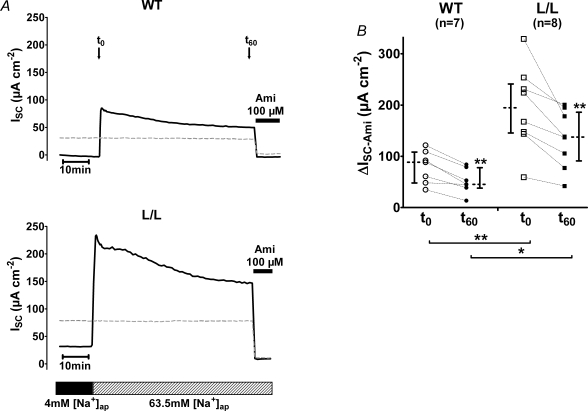
Figure 7. Sodium feedback inhibition is at least partially preserved in colon tissue of L/L mice.
The experimental protocol was similar to that described in Fig. 6. Tissue preparations were pre-incubated for 8 h using an apical low sodium (4 mm) bath solution. A, representative ISC traces from colon tissue of WT or L/L animals are shown. To investigate sodium feedback inhibition, ISC was monitored for 60 min after increasing the NaCl concentration in the apical bath solution to 63.5 mm (as indicated by the bar below the traces). ΔISC-Ami was estimated at time zero (t0) immediately after switching the apical bath solution from 4 mm to 63.5 mm NaCl and 60 min later (t60) when 10−4m amiloride (Ami) were applied as indicated by the black bar. The ISC traces, shown as dashed lines, represent control experiments in which colon tissues were pre-incubated with an apical bath solution containing 63.5 mm NaCl. Mock solution exchanges were performed at t0 to demonstrate that a solution exchange per se has no significant effect on ISC. B, summary of experiments as shown in A. Each circle or square represents an individual ΔISC-Ami value estimated at time zero (t0; open symbols) or after 60 min (t60; filled symbols) in colon tissue from WT (circles) or L/L (squares) animals. ΔISC-Ami values measured in matched tissues from the same animal are connected by dotted lines. In addition to the individual values, medians with interquartiles are shown.

