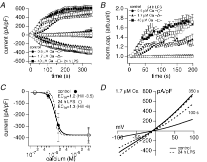
Figure 4. Calcium-activated (TRPM4-like) current.
A, average inward and outward currents (in pA pF−1) measured for 350 s, and B, normalized cell capacitance measured for 200 s after break-in from resting (filled) and activated (24 h 1 μg ml−1 LPS, open) microglial cells with 0.6 μm (circles, n = 7/5, ± s.e.m.), 1.7 μm (triangles, n = 13/10, ± s.e.m.) and 40 μm (squares, n = 19/7, ± s.e.m.) free calcium ([Ca2+]i) in the patch pipette. C, plateau inward currents (in pA pF−1) after 350 s versus log [Ca2+]i of resting (•) and activated (24 h 1 μg ml−1 LPS, ^) microglial cells. The sigmoidal fits of the inward current amplitudes revealed an EC50 of 1.2 μm Ca2+ (Hill coefficient =−3.5) for resting and 1.3 μm Ca2+ (Hill =−6) for activated microglial cells. D, average current–voltage relationships (I–Vs) of whole-cell currents (in pA pF−1) activated by 1.7 μm internal Ca2+ from resting (control, continuous traces, n = 9) and activated (24 h 1 μg ml−1 LPS, dotted traces, n = 9) microglial cells at 100 s and 350 s into the experiment.

