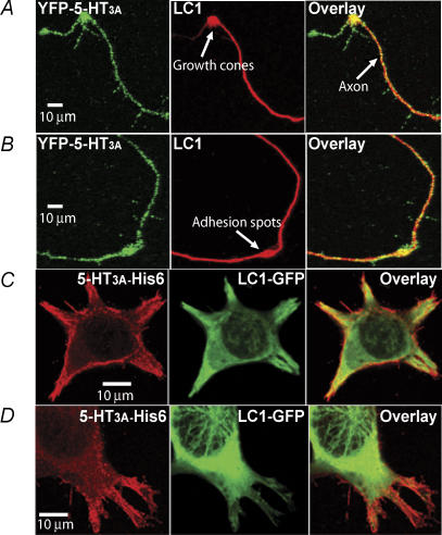
Figure 2. Colocalization of 5-HT3A receptors with LC1.
A and B, different examples of imaging illustrating colocalization of 5-HT3A receptors and LC1 in neurons. Hippocampal neurons were cultured in a serum-free medium for 4 days and transfected with the cDNA of the YFP-5-HT3A receptor. C and D, colocalization of 5-HT3A receptors and LC1 in HEK 293 cells. 5-HT3A-His6-Flag and LC1-GFP were cotransfected into HEK 293 cells. The red is the labelling of 5-HT3A receptor protein with anti-Flag-M2-Ab/anti-AlexaFluor 568. The green is the fluorescence from LC1-GFP.

