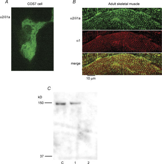
Figure 1. Identification of α2/δ1a isoform by a polyclonal antibody.
A, COS7 cells were transfected with a plasmid containing the full sequence of the α2/δ1a subunit, kindly provided by Drs N. Klugbauer and F. Hofmann and fixed 48 h later. Cells were examined for expression of the α2/δ1a protein by labelling with the α2/δ1 affinity-purified anti-1a isoform primary antibody (1 : 500) and Alexa-Fluor 488 goat anti-rabbit secondary antibody (1 : 1000). Confocal microscopy revealed diffuse staining of transfected cells. Control, untransfected, cells did not show any labelling. B, the tibialis anterior muscle was isolated from adult mice, frozen in liquid nitrogen, and sectioned. Sections of tissue were simultaneously labelled with the α2/δ1 affinity-purified anti-1a isoform antibody (1 : 500) and the α1 monoclonal antibody mAb 1A (1 : 1000; Morton & Froehner, 1987). The secondary antibodies were Alexa-Fluor 488 goat anti-rabbit and Alexa-Fluor 555 goat anti-mouse, both at 1 : 1000 (Molecular Probes). Examination of the sections with confocal microsopy showed co-localization of α2/δ1a and α1 labelling in a striated pattern, as would be expected for adult skeletal muscle. Note that α2/δ1a is the only isoform expressed in adult skeletal muscle. C, Western blot of whole-cell homogenate probed with the α2/δ1a polyclonal antibody. Two-day skeletal myotubes (C), COS7 cells transfected with α2/δ1a plasmid (1), and naïve COS7 cells (2). The affinity-purified antibody specifically recognizes both expressed and native α2/δ1a subunits. There was no signal in naïve COS7 cells.

