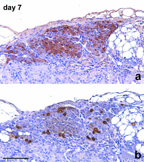
Fig. 10.
Consecutive sections of islets re-aggregated in a cluster. A large number of cells show positive staining for insulin (a). Also, the peripheral layer of the cluster is largely represented by B cells. Some glucagon-stained A cells are singly scattered peripherally and also in the inner part of the cluster (b). Immunoperoxidase stain for (a) insulin, (b) glucagon, 200×, scale bar = 150 µm.

