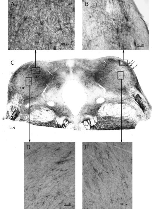
Fig. 5.
Distribution patterns of WFA binding sites in the IC of the rhesus monkey at Bregma −21.75 mm. DCIC, dorsal cortex of the IC; LLN, lateral lemniscal nucleus; ECIC, external cortex of the IC; CIC, central nucleus of the IC; open arrows indicate clusters of very large neurons with intensely stained WFA binding sites (B); windows in the CIC indicate examples of sheets of small neurons surrounded by faintly labelled perineuronal nets (D,E).

