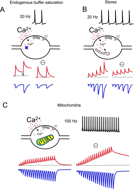
Fig. 1.
Mechanisms of control of presynaptic Ca2+ dynamics (I). For all the panels in this figure and Fig. 2 a general idealistic illustration of each of the mechanisms described in the text is shown. The stimulating protocols at which these mechanisms occur are depicted on top of each panel. Red traces represent free Ca2+ concentration. Blue traces represent postsynaptic responses. The hypothetical blockade of each mechanism is indicated with a minus symbol (‘–’). All panels represent general concepts and are not intended to show quantitative data. For an accurate idea of a realistic view of free Ca2+ dynamics and postsynaptic responses see Fig. 3. Only non-depressing synapses with a low release probability are considered. (A) The role of endogenous buffers in paired pulse facilitation (PPF) at a synaptic terminal. Two APs at 20 Hz produce two consecutive increases in Ca2+ concentration inside the terminal. The endogenous presynaptic Ca2+ buffers rapidly trap Ca2+ ions and can be partially saturated after the first AP at relatively high frequencies. Therefore, the residual Ca2+ when the second AP occurs is higher (left red trace), and this enhances transmitter release (left blue trace). The effect on free Ca2+ and EPSCs of a hypothetic blockade of buffer saturation is also illustrated. (B) Upon repetitive stimulation at 20 Hz, Ca2+ release from presynaptic Ca2+ stores can occur, contributing to the global Ca2+ signal. Through this mechanism neurotransmitter release is facilitated. The blockade of Ca2+ stores should reduce facilitation (only partially due to the presence of mechanism A) and increase resting Ca2+. (C) Intense (100 Hz) stimulation recruits presynaptic mitochondria and contributes to buffer the excess of Ca2+. In the absence of mitochondria the presynaptic Ca2+ concentration would reach higher levels during such a repetitive high-frequency stimulation, increasing the release rate.

