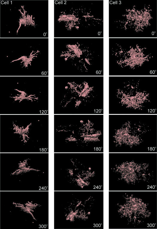
Fig. 2.
Surface rendering of GFAP/EGFP astrocytes in situ. An example of surface rendering of three GFAP/EGFP astrocytes in brain slices. For 3D reconstruction, the same images filtered using convolution filters were used as for morphometric measurements. The rotation of the cells to a specific angle is indicated in each image.

