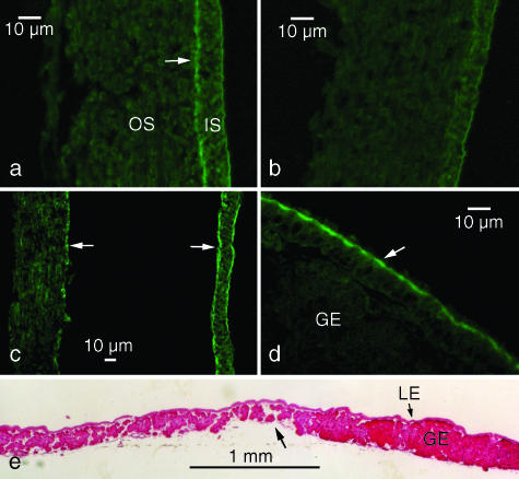
Fig. 8.
Histochemistry of shell membranes and the oviductal isthmus. (a–d) The antiserum stained a medial layer (arrow in a) between the inner (IS) and outer shell membrane (OS) at the blunt end in a uterine egg. This layer was not present in any other region of the shell membrane (b). In an oviposited egg, the medial layer was divided into two (arrows in c). The antiserum stained an apical region of the luminal epithelium of the isthmus (arrow in d). Immunofluorescent staining. (e) A sagittal section of the luminal epithelium (LE) showed that the glandular epithelium (GE) is poorly developed at the boundary (arrow) between the anterior (to the left) and posterior (to the right) of the isthmus. Haematoxylin-eosin staining.

