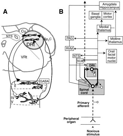
Fig. 1.
Pain facilitating circuits centred in the DRt. (A) Location of the DRt in a coronal section of the medulla oblongata obtained at 5.3 mm caudal to the interaural line (Paxinos & Watson, 1998) depicting the spino-DRt-spinal loops. The buffering role of GABAergic spinal neurons in counteracting the amplifying effect of the loop is included. (B) Interactions of the DRt, not only with the spinal cord, but also with other brain structures, namely the components of the pain control system (PAG, RVM, NTS and cVLM). The bold thick lines represent the circuit involved in signal amplification; dotted lines represent the circuit mediating aversive pain reactions; thin solid lines represent the circuit involved in counteracting signal amplification; dashed lines represent the signal transmission pathway. Filled neurons/arrows: excitatory actions; empty neurons/arrows: inhibitory actions. Adapted from Lima & Almeida (2002). Abbreviations: cc, central canal; Cu, cuneate nucleus; cVLM, caudal ventrolateral medulla; DRt, dorsal reticular nucleus; NTS, nucleus tractus solitarius; PAG, periaqueductal grey; RVM, rostroventromedial medulla; Sp5C, spinal trigeminal nucleus, pars caudalis; VRt, ventral reticular nucleus.

