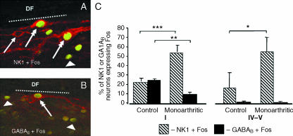
Fig. 3.
Effect of chronic pain in the nociceptive activation of spinal neurons bearing NK1 or GABAB receptors. (A,B) Confocal images of neurons double-immunostained for NK1 receptors and Fos protein (double arrows in A) or GABAB receptors and Fos protein (double arrow in A,B). The photomicrographs also depict cells labelled only for NK1 (arrow in A) or for the Fos protein (arrowheads) which was induced in response to noxious mechanical stimulation of the skin close to a chronically inflammed joint. The dotted lines mark the border between lamina I and the dorsal funiculus (DF). (C) Percentages of NK1- or GABAB-immunoreactive neurons in spinal laminae I or IV–V that express Fos in non-inflamed (control) or inflamed (monoarthritic) animals. Nociceptive activation of NK1-expressing neurons increases significantly in lamina I (P < 0.001) and IV–V (P < 0.05) whereas that of GABAB-expressing neurons decreases in lamina I (P < 0.01). Adapted from Castro et al. (2005).

