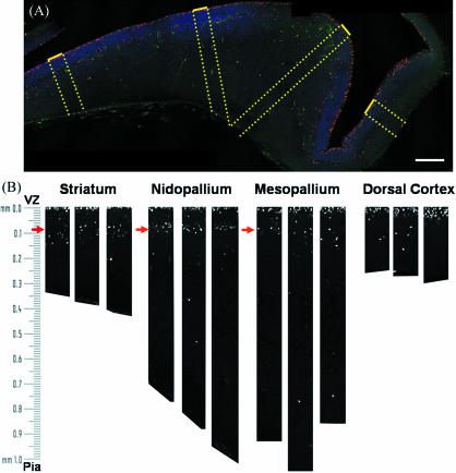
Fig. 5.
Reconstructions of the H3-immunoreactive nuclei in an E8 chick telencephalon. (A) Confocal microscopic reconstruction of a coronal section of an E8 chick brain. Phospho-histone H3-labelled cells (red) were counted in 100-µm bands running perpendicular from the VZ (dotted yellow lines). Nuclei are labelled with bisbenzimide (blue) and are most dense at the VZ. The outline of the section can be observed in the green channel. Bar, 200 µm. (B) Phospho-histone H3-positive nuclei were counted in three 100-µm bands from each region. Note that H3-labelled cells (white) cluster in the VZ (within 20 µm of the ventricle) and in the SVZ starting around 90 µm from the ventricle (red arrows).

