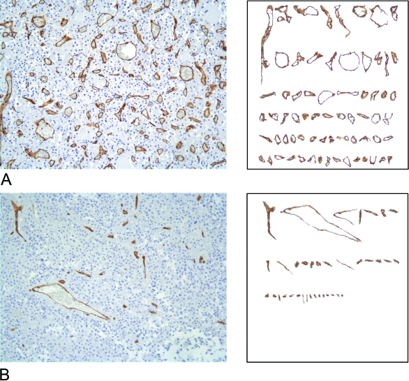
Fig. 1.
Examples of the histomorphological complexity of pituitary vascularity. Two-dimensional sections of normal (A) and adenomatous pituitary gland tissue (B) stained with antibodies raised against CD34 that specifically react with vessels by a standardised immunohistochemical procedure. A vessel catalogue is subsequently created for each digitised section: the immunopositive vessels are selected on the basis of the similarity of their colour on the RGB scale using a computer-aided image analysis system, and ordered on the basis of their magnitude. The highly variable nature of the vessel shapes and surfaces can be clearly seen.

