Fig. 4.
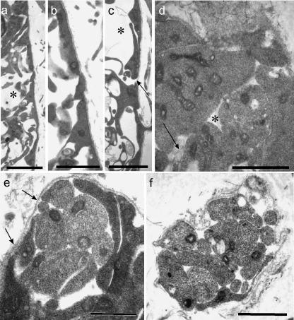
Electron micrographs of E11 rat neural tube at presumptive MEP loci. (a–c) Transverse sections of the neural tube show that the end-feet of glial processes comprise the presumptive glia limitans and form a tenuous or incomplete layer (c, arrow), which is covered by a continuous basal lamina (b, c); the underlying presumptive white matter contains prominent spaces (a, c asterisks), some of which contain flocculent material. (d) Tangential section through the glia limitans at E11; the glial end-feet contain prominent mitochondria; gaps (asterisk) are evident between them, containing in places folds of material resembling basal lamina (arrow). (e, f) Tangential sections parallel to the neural tube surface, showing: (e) a transversely sectioned early rootlet consisting of a few axons emerging through the glia limitans, surrounded by dark, loosely arranged glial end-feet, and also the continuity of the basal lamina covering the neural tube and rootlet (arrows); (f) a transversely sectioned early rootlet distal to the neural tube surface, consisting mostly of bare axons, with an extensive covering of basal lamina and an attenuated glial process apposed to part of its surface (below). Scale bars, a: 5 µm; b: 2.5 µm; c: 2 µm; d: 1.5 µm; e, f: 1 µm.

