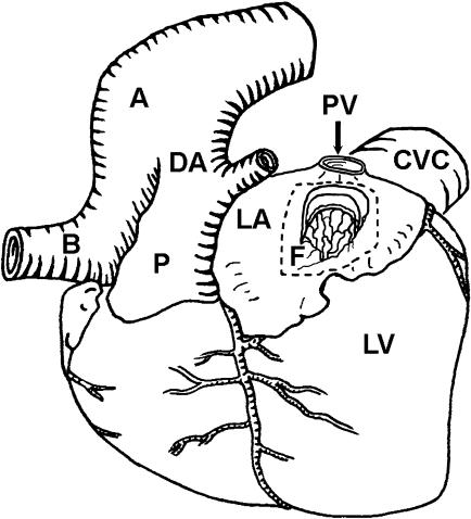
Fig. 1.
Schematic diagram of the heart of a cetacean fetus viewed from the left with a window cut into the left atrium. The relative positions of the aorta (A), left common carotid artery (B), ductus arteriosus (DA), pulmonary trunk (P), left atrium (LA), left ventricle (LV), caudal vena cava (CVC), pulmonary vein (PV) and the fold of tissue (F) representing the developed septum primum lying in the lumen of the left atrium are indicated. The latter is distal to the foramen ovale, which it obscures. Pericardial, pleural and lung tissues have been removed.

