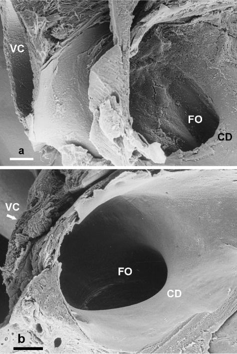
Fig. 2.
(a) Scanning electron micrograph of the foramen ovale (FO) in a white whale (Delphinapterus leucas) fetus viewed from the right atrium, illustrating the curved edge of the crista dividens (CD) of the interatrial septum, and the caudal vena cava (VC). Scale bar = 1 mm. (b) Scanning electron micrograph of the foramen ovale (FO) in a humpback whale (Megaptera novaeangliae) fetus viewed from the right atrium, illustrating the curved edge of the crista dividens (CD) of the interatrial septum and the caudal vena cava (VC). Scale bar = 1 mm.

