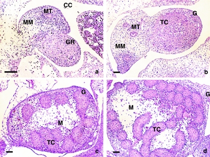
Fig. 1.
Morphological stages of testis development: (a) E12 embryo presenting morphologically undifferentiated gonads; (b) E13 embryo showing the developing testis; (c,d) E14 and E15 embryos presenting differentiated testicular cords. H&E staining. CC, coelomic cavity; G, gonad; GR, gonadal ridges; MT, mesonephric tubule; MM, mesonephric mesenchyme; M, mesenchyme of testis; TC, testicular cords. Scale bar, 50 µm.

