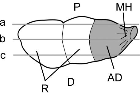
Fig. 1.
Diagrammatic representation of the articular disc (AD) and meniscal homologue (MH), seen from the carpal side. This shows the location of the sections taken from the palmar (a), central (b) and dorsal (c) regions of the TFCC for immunohistochemistry. Note that the disc and meniscal homologue cover the lower end of the ulna. D, dorsal aspect; P, palmar aspect; R, radius.

