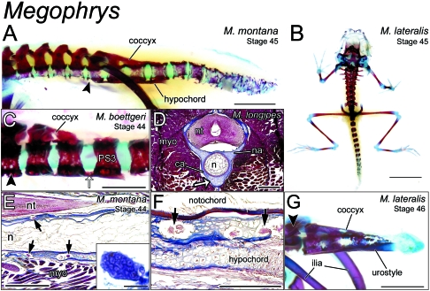
Fig. 3.
Remodelling of the postsacral skeleton of Megophrys at metamorphosis. (A) Lateral view of the axial skeleton (Vertebrae I–III not shown) of a Stage-25 Megophrys montana metamorph (CAS 138410). With the exception of the sacrum (black arrowhead) and those postsacral vertebrae in contact with the hypochord and coccyx, centra are fragmented and porous along the length of the vertebral column, suggestive of osteoclastic degradation. Scale bar, 2 mm. (B) A cleared-and-stained metamorph of Megophrys lateralis (Stage 45; ROM 42366) in dorsal perspective. Proximal postsacral centra are perichordal, whereas, more distally, centra comprise discrete ossification centres. Scale bar, 4 mm. (C) Close-up lateral view of the immediately postsacral vertebrae of a Megophrys boettgeri metamorph (Stage 44; A-30639.1). Postsacral Vertebrae 1–3 are joined ventrally by a longitudinal bridge (white arrow), which extends from the caudal margin of PS3. This structure will go on to form the hypochord in later stages. The dorsal surface of PS1 and 2 are fused as the coccyx. The hindlimb skeleton has been disarticulated from the sacrum (arrowhead), allowing for an unobstructed view. Scale bar, 1 mm. (D) Transverse cross-section at the level of PS1 of a Stage-31 tadpole of Megophrys longipes. The hypochordal ridge (white arrow) is clearly continuous with the perichordal tissue constituting the centrum (ca). myo, myotome; n, notochord; na, neural arch; nt, neural tube. Scale bar, 0.5 mm. (E) Parasagittal section through the sacral region of an M. montana metamorph (Stage 44; CAS 138409.2), showing multinucleate, osteoclast-like cells (arrows) embedded in peri-notochordal osseous tissue. Scale bar, 0.5 mm. Inset shows a close-up view of a multinucleate osteoclast in section. Scale bar, 50 µm. n, notochord; nt, neural tube; myo, myomere. (F) Sagittal section through the same specimen in E. Osteoclast-like cells are clearly visible in the peri-notochordal osseous tissue, but absent from the nascent hypochord, lying ventral. Scale bar, 0.25 mm. (G) Lateral view of the sacrum (black arrowhead) and caudal skeleton of a Stage-46 M. lateralis (ROM 42368) nearing the very end of metamorphosis. Postsacral Vertebrae 1–3 have been incorporated into the urostyle. All other supernumerary centra have been resorbed by this point. Scale bar, 2 mm.

