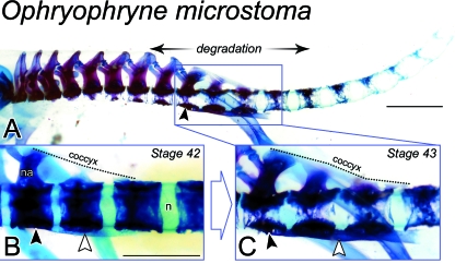
Fig. 4.
Remodelling of the postsacral skeleton of Ophryophryne microstoma at metamorphosis. (A) The postcranial skeleton of a Stage-43 metamorph (ROM 42345) shown in lateral perspective. Vertebral centra appear more degraded at distal levels relative to the sacrum (black arrowhead). The most rostral presacral centra are completely degraded at their lateral and ventral faces, consistent with the epichordal type described by Griffiths (1963). More caudally, including all postsacral vertebrae, centra are noticeably porous, but still encompass the notochord (i.e. perichordal type). Scale bar, 1.5 mm. (B) Lateral view of the sacrum (black arrowhead) and PS1–3 of a Stage-42 metamorph (ROM 42344). Dorsally, the coccyx (traced in broken line) joins the sacrum, PS1 and PS2. The hypochord (white arrowhead) can be clearly seen ventrally, spanning the sacrum to PS2. Vertebral centra enclose the notochord (n) and are not degraded. na, neural arch. Scale bar, 1 mm. (C) By Stage 43, presented here as a close-up of the sacral region of A, centra are degraded laterally and the coccyx (broken line) and hypochord (white arrowhead) have each thickened and now span the sacrum (black arrowhead) to PS3.

