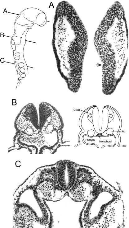
Fig. 2.
Neural crest at stage 10. Alum cochineal. (A) Mesencephalic crest from the neural folds can be seen above. The crest cells are darker and more rounded than those of the mesoderm. The optic sulcus (arrow) is visible and the optic primordium is present. (B) Facial crest in stage 10 is emerging laterad from the wall of the neural groove. (C) Hypoglossal crest is migrating between the neural tube and the first somite. The planes of section are shown in the key. Figure 2B is taken from R. O’Rahilly and F. Müller, The Embryonic Human Brain, 3rd edn. Copyright ©, 2006, Wiley-Liss. Reprinted by permission of John Wiley and Sons, Inc.

