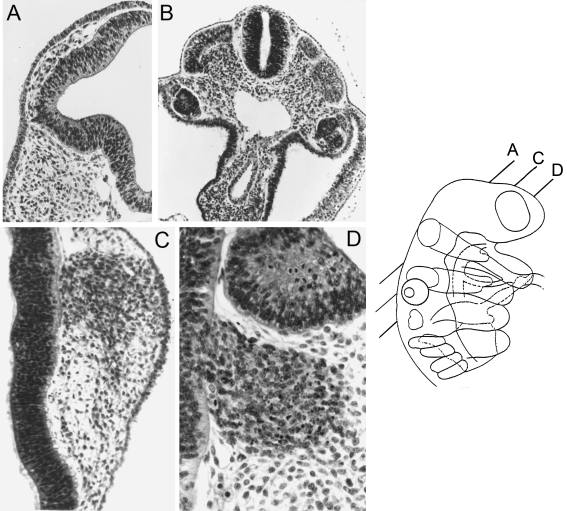
Fig. 4.
The neural crest at stage 12. The planes of sections of A, C, and D are shown in the key. Alum cochineal/haematoxylin and eosin. (A) Optic crest is clearly emigrating from the optic vesicle. (B) Spinal crest in the thoracic region. The roof cells of the neural tube resemble the crest cells that are visible between the tube and the dermatomyotomes. (C) The trigeminal ganglion is connected here to the surface ectoderm by cells believed to be emigrating from the latter. (D) The facial ganglion (seen here below the otic primordium) of the embryo shown in C. The cellular columns of the neural wall continue into the ganglion and a basement membrane is absent at this level.

