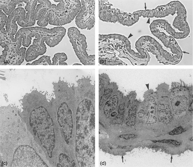
Fig. 1.
Fimbriae and bursa. (a) Semithin section through fimbrial folds at metestrus stage covered by ciliated and non-ciliated cells. ×245. (b) Semithin section through the bursa at estrus stage. Arrowheads, bursa epithelium with ciliated and non-ciliated cells; arrows, serosal epithelium. ×245. (c) Electronmicrograph of fimbrial epithelium at metestrus, depicting only non-ciliated cells. ×6100. (d) Bursa wall, with cuboidal bursal epithelium (arrowhead) and flat serosal epithelium (arrows) at metestrus. ×2300.

