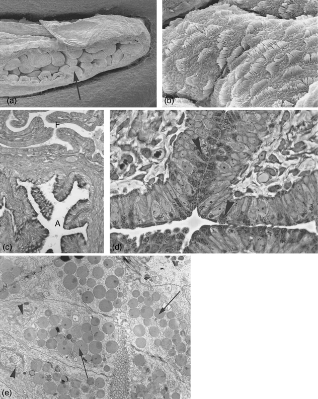
Fig. 2.
Ampulla at estrus. (a) Scanning electron microscopy. Overview of ampullar folds (arrow). ×560. (b) Folds covered with an epithelium consisting of ciliated and non-ciliated, secretory cells. ×7000. (c) Light microscopical section stained with PAS. Note the intensive positive reaction of the secretory cells in the ampulla (A) and the negative reaction of the fimbrial epithelium (F) where no staining products could be detected. ×145. (d) Semithin section with ampullar epithelium exhibiting non-ciliated, secretory cells, densely packed with secretory granules (arrowheads) at estrus. ×615. (e) Electronmicrograph of secretory cells at estrus, filled with secretory granules often of heterogeneous density (arrows) and highly active Golgi complexes (arrowheads). ×6400.

