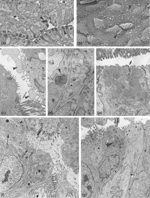
Fig. 3.
Ampulla at post-and metestrus. (a) Semithin section through ampullar epithelium at postestrus stage. The number of secretory active cells (arrowheads) and the amount of secretory granules per cell are reduced. ×500. (b) Scanning electronmicrograph showing postestrus stage with extruded secretory material (arrowheads), partially membrane bound after apocrine secretion. ×3200. (c–g) Electronmicrographs. (c) Non-ciliated cell with apocrine cytoplasmic exocytosis (arrowheads). ×3900. (d) Secretory products in degradation in form of whorls (arrowhead). ×4700. (e) Section through a secretory cell at postestrus still hosting numerous secretory granules, one cell carrying a solitary cilium (arrow). ×8000. (f) Some of the non-ciliated cells show apically numerous translucent small vesicles (arrows) beside few secretory granules. ×6400. (g) Ampullar epithelium at metestrus, occasionally a few remnant secretory granules are left (arrowhead). ×5000.

