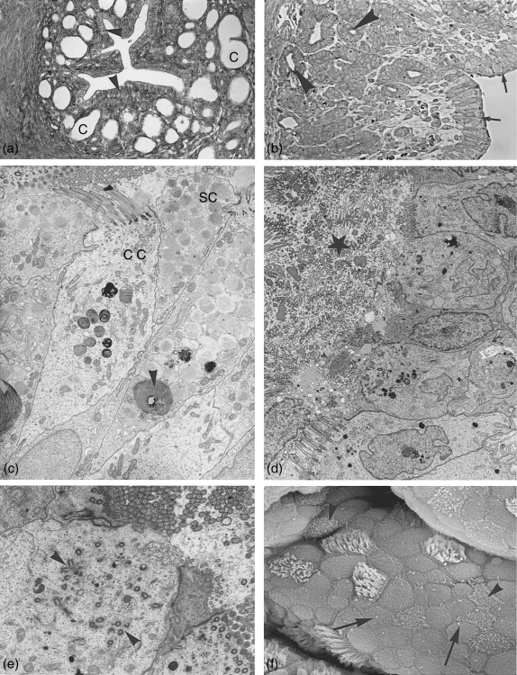
Fig. 4.
Isthmus. (a) Light microscopy: cross section through the isthmus region close to the uterus showing surface epithelium (arrowheads) and crypts (C). ×165. (b) Semithin section through the isthmus region with surface epithelium (arrows) and crypts (arrowheads). ×250. (c–f) Isthmus surface epithelium. (c) Electron microcoscope pictures of isthmus surface epithelium at postestrus stage with ciliated cell (cc) and non-ciliated secretory cells (sc) still containing numerous secretory granules. Degeneration processes (whorls) have started (arrowheads). ×4500. (d) Lumen at metestrus filled with massive deciliation and secretion products (star). Hardly any secretory granules are left in the secretory cells. ×3300. (e) At the same time ciliogenesis has started. Note the multiplication of basal bodies moving to the apical surface (arrowheads). ×6900. (f) Scanning electron microscope: isthmus surface at late metestrus/early proestrus showing numerous non-ciliated cells with solitary cilia (arrows) and ciliated cells exhibiting neo-ciliogenesis at different stages (arrowheads). ×7200.

