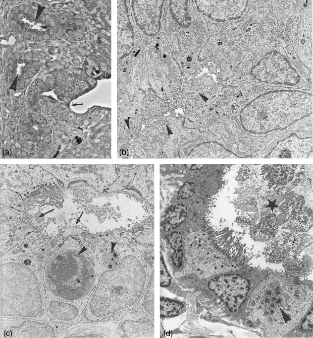
Fig. 5.
Isthmus crypts. (a) Semithin section, invagination of the surface epithelium (arrow) into crypts. Crypt epithelium consists at estrus mainly of secretory cells with bulging apices (arrowheads). ×310. (b) Electron microscopy: crypt cells at estrus. Secretion products exist in numerous translucent vesicles located in bulging cell apices (arrowheads). ×6120. (c) Crypt cells at late postestrus shedding apices and cilia (arrows). Small secretory vesicles can still be found in individual cell apices. Lysosomes and aptotosic cells are typical (arrowheads). ×4200. (d) Crypt at metestrus filled with shed cilia (star), cell with signs of neo-ciliogenesis (arrowhead). ×4300.

