Fig. 6.
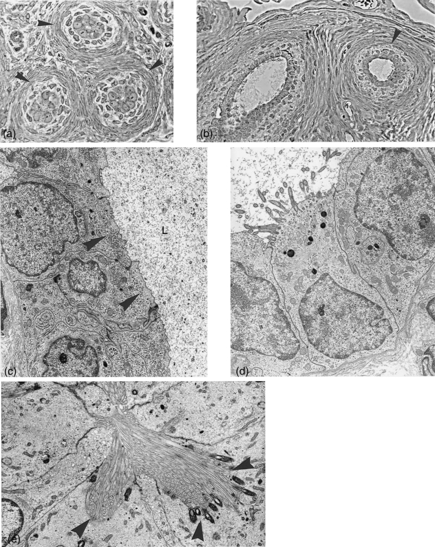
Epoophoron. (a) Cross section (semithin) through epoophoron tubules with narrow lumen and distinct connective tissue layers (arrowheads) surrounding the epithelium. ×450. (b) Semithin sections through tubules with wider lumina lined with ciliated and non-ciliated cells. Both figures are taken from the same animal at metestrus stage. ×300. (c) Electron microscopy: non-ciliated epithelium lining the epoophoron tubules at metestrus, with numerous small vesicles at the cell apices (arrowheads) and secretion product in the lumen (L). ×6900. (d) A ciliated cell of the same tubule. ×6050. (e) A different type of ciliated cell, where cilia sit in deep invaginations with pronounced basal bodies (arrowheads). ×7100.

