Fig. 4.
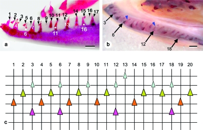
Tooth pattern in marine animals. (a) Female. Lateral view of the left dentary, with tooth positions numbered. Due to the anterior inward curvature of the dentary, position 3 appears to lie caudal to position 4. Note the retention of a tooth undergoing resorption in position 6, 11 and 16 (white asterisks). Bar = 1 mm. (b) Male. Dorsal view of the left dentary. The epithelial connection between functional teeth and their successors is well visible (blue arrowheads). Functional teeth are located labially, young replacement teeth lingually. Arrows indicate old functional teeth that are retained in the presence of young replacement teeth (positions 3, 6, 12 and 18). Bar = 1 mm. (c) Graphical representation of the dentition on the dentary of marine animals, as exemplified by the left dentary of the animal shown in Fig. 4b. Vertical lines represent (numbered) tooth positions. Horizontal lines connect teeth in a similar stage of development (for replacement teeth) or functionality (for functional teeth). Lingual to the top, labial to the bottom of the chart. Four stages of tooth development can be distinguished on cleared and stained animals: young replacement teeth (small light green triangles), advanced replacement teeth (large dark green triangles), functional teeth (whether newly attached or mature) (large orange triangles), and functional teeth undergoing resorption (large purple triangles). Note that sets of three teeth in descending order of development succeed each other along the tooth row (positions 1–3, 4–6, etc.). This pattern is interrupted by an extra tooth germ in position 13, and an apparent loss of a tooth between positions 16 and 17. Due to the retention of a functional tooth together with a young replacement tooth (positions 3, 6, 12 and 18), functional teeth appear to be irregularly distributed and separated by variable numbers of replacement teeth.

