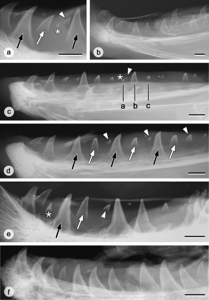
Fig. 5.
Tooth pattern on the left lower jaw in grilse, salmon and kelts. (a) Characteristics of teeth on X-rays as exemplified by a male grilse. A tooth germ in formation (white arrowhead) can be clearly distinguished from the remnants of a functional tooth in resorption (white asterisk): tooth tips in resorption are more heavily mineralised and they do not possess a V-shaped edge to the dentine as in developing teeth. Attached functional teeth are indicated by black arrows, an advanced replacement tooth by a white arrow. Bar = 10 mm. (b) X-ray of the left dentary of a male grilse. The graphical representation of the tooth pattern corresponding to this dentary is shown in Fig. 6a. Bar = 10 mm. (c) X-ray of the left dentary of a female grilse. In this animal, more posterior positions appear to be advanced with respect to anterior positions. The three black lines (a, b, c) indicate three consecutive positions that, if the pattern were continued from the front, would have been constituted by a functional tooth, a more advanced and a younger replacement tooth. Instead, the functional tooth has undergone resorption, leaving only the tooth tip (white asterisk), together with an early replacement tooth (white arrowhead). The next positions are similarly advanced. Together this creates the impression that, more anteriorly, four instead of three adjacent positions show decreasing developmental stages. Bar = 10 mm. (d) X-ray of the left dentary of a female salmon. Note the repetition of functional teeth (black arrows), advanced replacement teeth (white arrows), and young replacement teeth (white arrowheads) along the row. Bar = 10 mm. (e) X-ray of the left dentary of a male kelt. A pattern of three consecutive teeth of diminishing developmental stage is still recognizable in some parts of the tooth row (cf. black arrow, white arrow, white arrowhead); elsewhere adjacent teeth have attached. An asterisk indicates the tip of a tooth in resorption. Bar = 10 mm. (f) X-ray of the left dentary of a female kelt. Note that almost all teeth are attached; developing replacement teeth are rare. Bar = 10 mm.

