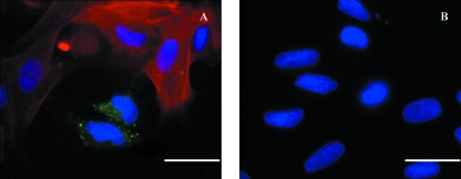
Fig. 7.
Coculture preparations with dual-label staining directed to 5HT3 receptors (green) and µ-smooth muscle actin (red) antibodies for characterization of neurones and ISMC respectively. DAPI mount was used for labeling cell nucleus (blue). Positive diffusion of the 5HT3 receptors antibody within the neuronal cell bodies opposed by neighbouring cells identified as ISMC (A). Negative controls showed no staining to both primary antibodies (B). Scale bar = 25 µm.

