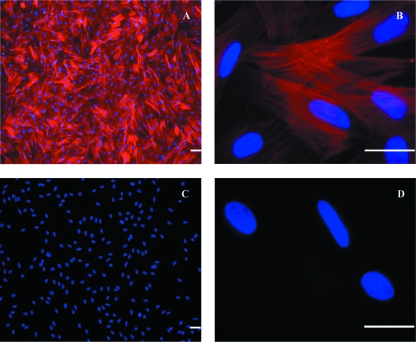
Fig. 8.
ISMC preparations with dual-label staining directed to 5HT3 receptors (green) and α-smooth muscle actin (red) antibodies for confirmation of the nature of the purified ISMC. DAPI mount was used for labeling cell nucleus (blue). Uniform ISMC actin filaments stained positive throughout the culture (A,B). Negative labeling to 5HT3 receptors antibody confirmed the absence of the neurones in the preparation (A,B). Negative controls showed no staining to both primary antibodies (C,D). Scale bar = 25 µm.

