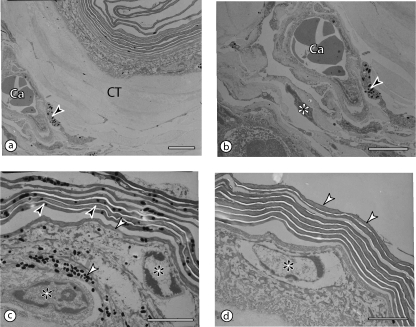
Fig. 5.
Transmission electron micrographs of the wing web, showing the layers and the position of the core capillaries. All scale bars = 3 µm. (a,b) The capillaries (Ca) are placed within the core tissue (CT), approximately equidistant from both surfaces. Notice a melanocyte (arrowhead in b) with melanin granules and a fibroblast (asterisk) close to the capillary. (c,d) The non-keratinized part of the epidermis is one to two cells thick (asterisks) and is covered by the stratum corneum with about 10 layers of flattened keratinocytes (dark arrowheads in c; white arrowheads in d). Both the keratinocytes and the other epidermal cells may contain melanin granules (white arrowheads in c).

