Fig. 4.
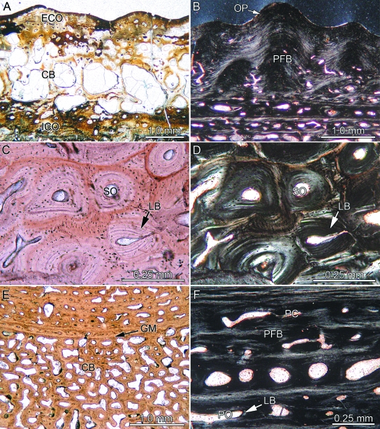
Bone histology of temnospondyl amphibians. (A) Complete section of Trimerorhachis sp. (TMM 40031-59) showing interior cancellous bone (CB) framed by external and internal cortical bone (ECO, ICO). (B) Close-up of external ornamentation pattern (OP) and parallel-fibered bone (PFB) of Gerrothorax pustuloglomeratus (SMNS 91012) in polarized light. Close-up of the cancellous bone of external cortex of Mastodonsaurus giganteus (SMNS 91011; transverse section) in (C) normal and (D) polarized light. Note lamellar bone (LB) of older and younger generations of secondary osteons (SO) forming Haversian bone. (E) Close-up of cancellous bone of G. pustuloglomeratus (SMNS 91012; transverse section) showing a compact primary growth mark (GM). (F) Close-up of internal cortex of former specimen (transverse section) strongly vascularized by primary osteons (PO, lined with LB) and primary vascular canals (PC).

