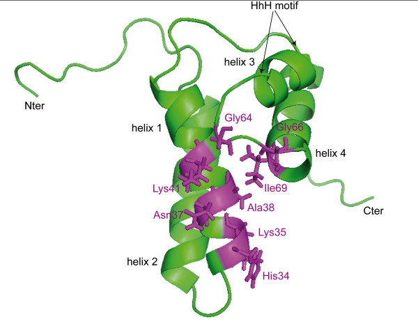
Figure 1.
Mapping residues found close to pamoic acid by docking experiments on the structure of the 8 kDa domain. Ribbon view of the 3D structure of the 8 kDa domain (PDB code 1DK3), highlighting residues found to be involved in binding with pamoic acid from docking experiments (colored in magenta). The four alpha helices and the "HhH" motif are annotated. Picture was prepared with PyMOL [48].

