
Figure 4.
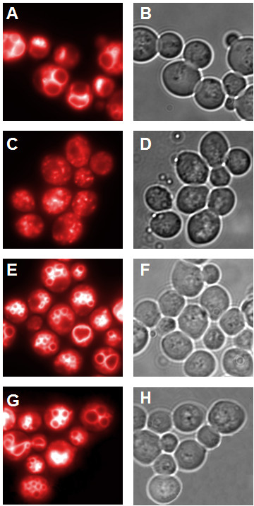
Vacuolar morphology. A,C,E,G: fluorescence microscopy; B,D,F,H: light microscopy; A,B: CG781 cells, C,D: CG783 cells, E,F:CG783 pRS314 Vam7 transformed cells, G,H: CG783 pRS314 Vam7-TB transformed cells (magnification 100×).
Official websites use .gov
A
.gov website belongs to an official
government organization in the United States.
Secure .gov websites use HTTPS
A lock (
) or https:// means you've safely
connected to the .gov website. Share sensitive
information only on official, secure websites.
Vacuolar morphology. A,C,E,G: fluorescence microscopy; B,D,F,H: light microscopy; A,B: CG781 cells, C,D: CG783 cells, E,F:CG783 pRS314 Vam7 transformed cells, G,H: CG783 pRS314 Vam7-TB transformed cells (magnification 100×).