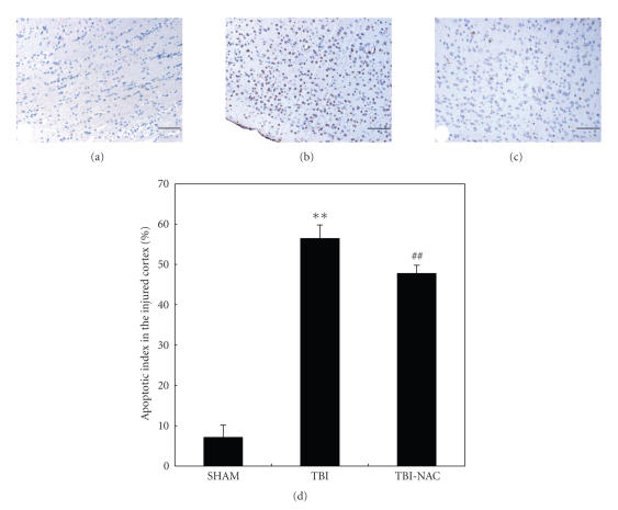
Figure 7.
TUNEL immunohistochemistry staining of the injured cortex in SHAM group (n = 5), TBI group (n = 6), and TBI-NAC group (n = 6). (a) SHAM group rats showing few TUNEL apoptotic cells. (b) TBI group rats showing more TUNEL apoptotic cells stained as brown. (c) TBI-NAC group rats showing less TUNEL apoptotic cells than TBI group (scar bar, 50 μm). (d) Administration of NAC significantly decreased the apoptotic index in rat injured brain following TBI. **P < .01 versus SHAM group; ## P < .01 versus TBI group.

