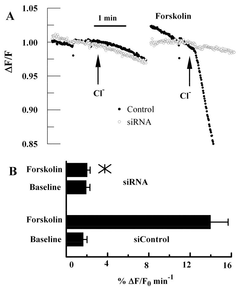
Figure 2.
Apical Cl- Permeability in control and CFTR siRNA treated cells. Cells were depleted of Cl-, loaded with the halide sensitive fluorescent dye MEQ and perfused on basolateral and apical sides with Cl- free ringer’s solution. Relative apical Cl- permeability is measured as the initial rate of MEQ fluorescence quenching upon addition of Cl- to the apical perfusing solution. A. Representative experiments showing the change in MEQ fluorescence relative to the starting fluorescence value in response to addition of chloride on the apical side in the absence and presence of 10 μM forskolin for siControl and CFTR siRNA treated cultures. B. Bar graph summarizes the Cl- permeability data (n=8). *, mean value significantly different compared to control (p<0.05); error bars show Standard Deviation.

