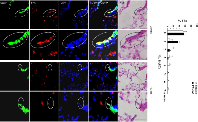
Figure 4. PX-866 treatment decreases BASC numbers in KrasLA1 mice.
Immunofluorescent staining to detect cells at terminal bronchi that co-express CCSP and SPC. Encircled BASCs in top panels (×10 magnification) illustrated in lower panels at higher magnification (×40). Bar graph indicates percentages of terminal bronchi with the indicated numbers of BASCS. Calibration bars in images of H&E stained tissues represent 50 μm.

