
Figure 2.
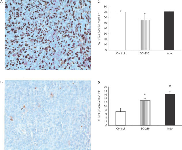
Apoptosis and proliferation in 4TI mammary fat pad tumours. (A) PCNA-positive (proliferating) cells within tumours (original magnification ×400). (B) TUNEL-positive (apoptotic) cells within tumours. Apoptotic cells stain brown after TUNEL staining (original magnification×400). (C) % PCNA positive cells in tumours from control mice and mice treated with SC-236 or indomethacin. Three high power fields per section (400×magnification (×40 objective and ×10 ocular)). Data represented as mean±s.e.m. P=n.s. (D) Number of TUNEL positive cells per h.p.f. Five high power fields per section (400×magnification (×40 objective and ×10 ocular)). SC-236 or indomethacin significantly increased apoptosis relative to tumours from control mice (one section from each of six mice per group). Data represented as mean±s.e.m. *P<0.05.