
Figure 2.
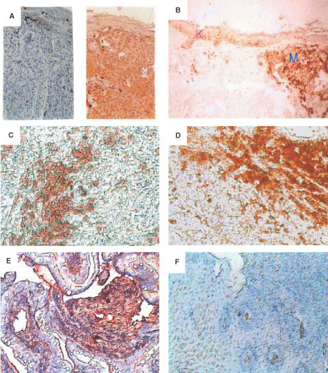
Immunohistochemical detection of MC1R on primary melanoma tissues, normal keratinocytes and metastatic melanoma tissues. Paraffin sections of primary melanoma tissues and normal skin tissues and metastatic melanoma tissues were stained with mAb MP1-1B7 or IgG control followed by a biotinylated anti-Ig mAb and developed as described in Materials and Methods. (A) normal naevus 10× (left) and primary melanoma (right) (B) Comparison of the intensity of labelling between a primary melanoma (M) and keratinocytes (K)×10. (C) Melanoma infiltrated lymph node (MP1-1B7)×10. (D) Maxilar metastasis (MP1-1B7)×10. (E) Intestinal metastatic melanoma (MP1-1B7)×10, and (F) Intestinal metastatic melanoma (Ig negative control)×10. In panels A, E, and F, nuclear staining was performed using haematoxylin solution.