
Figure 4.
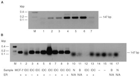
Occurrence of M41 mRNA in benign and malignant human mammary cell lines and breast tumour specimens by RT–PCR. Total RNA from human mammary cell lines. Normal breast derived Huma 7 (A, lane 1), benign breast derived Huma 123 (lane 2), Huma 109 (A, lane 3), and breast carcinoma-derived cell lines, MCF-7 (A, lane 4), T47-D (A, lane 5), ZR-75 (A, lane 6), MDA-MB-231 (A, lane 7), and total RNA from breast carcinoma specimens (B, lanes 2–9, 12–13), normal breast specimens (B, lanes 10 and 17), and benign breast specimens (B, lanes 11 and 16) were amplified by RT–PCR using primers specific for M41 yielding PCR products of 147 bp. (B), lanes 14 and 15 are the negative RT–PCR controls, and lane 1 is MCF-7 cell line RNA as positive control. The resulting RT–PCR products were subjected to agarose gel electrophoresis and stained with ethidium bromide, as described in Materials and Methods. Lane M, DNA molecular weight markers. The estrogen receptor status of the specimens is shown beneath panel B as +, positive or -, negative. N/A is not available. The faint bands in A, lanes 1–3 and 7 arise from the amplification of a low, otherwise undetectable, level of M41 mRNA in these specimens. The diffuse bands in B, lanes 2, and 10–17 at <100 bp are caused by primer dimers. DNA sequencing of the 147 bp band confirmed its identity with the expected sequence of the amplified band.